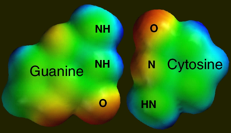
DNA provides the most important example of hydrogen-bonding. This figure was constructed from two separate models. It contains an electrostatic potential map of guanine (left) and a map of cytosine (right) oriented so that the two bases can engage in Watson-Crick base-pairing. The complementary nature of the intermolecular region is obvious. An electron-poor hydrogen on one molecule is positioned opposite an electron-rich atom on the other (this is a hydrogen bond!), and there are three such pairings.
These models not only make it easy to explain hydrogen-bonding, and they also make it possible to test student understanding of hydrogen-bonding concepts. For example, careful examination of the top NH in guanine shows that this hydrogen is more electron-poor (a better hydrogen-bond donor) than the middle NH in guanine. This is probably due to the fact that the top NH has an electron-poor neighbor, while the middle NH is sandwiched between electron-poor and electron-rich groups. Similarly, the bottom O in guanine is sandwiched between electron-poor and electron-rich groups, but it is more electron-rich on the side adjacent to the neighboring electron-rich group (bottom of molecule).
A simple follow-up question that can be directed at students is, "which type of base pair will stick together more strongly - one in which all of the donor groups are found on one base and all of the acceptor groups are found on the other base? or, one in which donor and acceptor groups are mixed between the two bases (like guanine and cytosine)?" Students can use the principles that have already been derived to answer this question, and they are surprised to find out that the "natural" base pair is not the strongest.